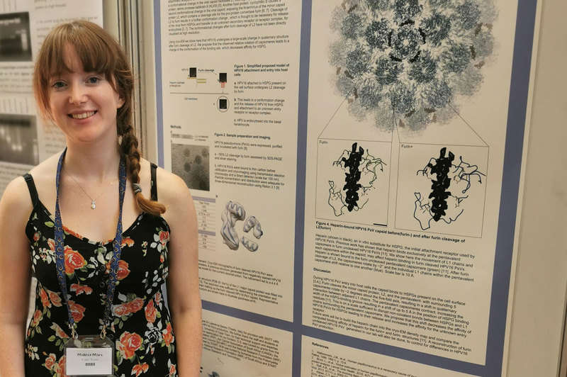
MSc student lauded for world-class HPV research
08 March 2021 | Story Niémah Davids. Photo Supplied. Voice Neliswa Sosibo. Read time 5 min.
Melissa Marx, an MSc student in the University of Cape Town’s (UCT) Faculty of Health Sciences (FHS), is conducting groundbreaking research in the field of cervical human papillomavirus (HPV). She hopes that her findings will set a strong foundation as scientists work towards discovering therapeutics to decrease the high burden of HPV infection in the country, on the continent and around globe.
Marx is a medical biochemistry student in UCT’s Department of Integrative Biomedical Sciences based in the FHS. Her research into HPV, which forms part of a larger department programme, was funded by the Global Challenges Research Fund Synchrotron Techniques for African Research and Technology (GCRF START).
Marx is among a long list of women at UCT and in South Africa who are making strides in the fields of science, technology, engineering and mathematics. GCRF START said that Marx’s research into HPV addresses a vital global challenge using world-class science and is in line with the United Nations Sustainable Development Goals.
“Without the help of the GCRF START grant, much of my research would not have been possible.”
“Without the help of the GCRF START grant, much of my research would not have been possible. The grant enabled me to apply cutting-edge structural biology techniques to gain insights into the structure of HPV,” she said.
“I was also incredibly fortunate to have collected data at the Electron Bio-Imaging Centre at Diamond Light Source in the United Kingdom. This was only possible with funding from the GCRF START grant.”
About HPV
HPV is a common viral infection of the reproductive tract and is spread through skin-to-skin contact. There are more than 100 different HPV types, which infect the genitals, mouth and throat.
According to the World Health Organization, most HPV infections clear on their own. However, there is a risk that the infection may become persistent and lead to lesions within infected areas. These pre-cancerous lesions may progress and develop into invasive cervical cancer. Cervical cancer is the fourth most common cancer among women worldwide. In 2018 roughly 570 000 cases were reported, and it represented about 7.5% of all female cancer deaths globally. Cervical cancer claims more than 311 000 lives every year, and 85% of these cases occur in low- to middle-income countries.
The research
Marx’s research focused on visualising the effect of an enzyme found within the reproductive tract on the structure of the virus, as it occurs during the infection process. To achieve this, she and fellow researchers studied HPV pseudoviruses (non-infectious and synthetic viruses) using laboratory-based techniques, structural biology and computational work.
The team closely studied the changes in the HPV structure as they occurred during infection, using high-resolution cryo-electron microscopy and three-dimensional reconstructions of the virus. During this process, the researchers completed the first high-resolution structural model of HPV, which occurred during the infection process, in an intermediate form. This detailed information on the structure of the virus allows greater insight into the HPV infection process and adds to scientific knowledge on the events that occur during infection.
Scientists hope to use the information and other related research to develop preventative and therapeutic options for HPV infection.
“HPV is the cause of most cervical cancer cases worldwide, and an urgent intervention is necessary to reduce the high burden of disease.”
Why HPV?
HPV is the cause of most cervical cancer cases worldwide, and an urgent intervention is necessary to reduce the high burden of disease. There are no effective treatments for HPV infection, but prophylactic vaccines are available globally to prevent infection in unexposed individuals.
While these vaccines are safe and effective, they are ineffective against existing infections and may not prevent infection by all HPV types. Going forward, Marx and her fellow researchers will continue to study the early-entry mechanisms of HPV during infection, using cryo-electron microscopy. This process will depict a detailed picture of the virus and how it operates. She said that scientists have a single aim: to use this and other data available to help them develop new treatment options to prevent the spread of HPV.
Marx said that UCT’s Electron Microscope Unit played an integral part in her research process. Researchers used the electron microscopes and cryo-EM preparation resources to assess the HPV samples, and thanks to these excellent resources, only the best samples were sent to the Electron Bio-Imaging Centre for scrutiny.
As Marx continues her research in earnest, additional findings will be published in a peer-reviewed journal in due course.
 This work is licensed under a Creative Commons Attribution-NoDerivatives 4.0 International License.
Please view the republishing articles page for more information.


