UCT hosts Nobel laureate for lecture on cryo-electron microscopy
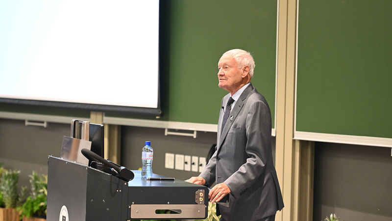
26 August 2024 | Story Stephen Langtry. Photos Lerato Maduna. Video Production Team Ruairi Abrahams, Boikhutso Ntsoko and Nomfundo Xolo. Read time 3 min.On Tuesday, 20 August, the University of Cape Town (UCT) hosted a Vice-Chancellor’s Open Lecture, which was delivered by Nobel laureate Professor Joachim Frank. The event, held at the New Lecture Theatre (NLT) on UCT’s upper campus, attracted a diverse audience who learned more about the revolutionary field of cryogenic electron microscopy (cryo-EM).
This was the first Vice-Chancellor’s Open Lecture for 2024. The lecture, titled “Cryo-electron Microscopy: A New Foundation for Molecular Medicine and Drug Design,” provided an exploration of how cryo-EM has transformed the study of biological molecules. In introducing the speaker, UCT’s vice-chancellor (VC), Professor Mosa Moshabela, said, “At UCT we have been developing the cryo-EM capability for more than 20 years now in the Electron Microscope Unit”.
Professor Frank, a pioneer in this field, shared insights into how this cutting-edge technology has enabled researchers to determine the structure of molecules in unprecedented detail. He emphasised how the ability to observe molecules freely suspended in solution, known as “single particles”, has paved the way for significant advancements in understanding molecular interactions within cells.
Frank detailed the process of cryo-EM, which involves freezing samples by plunging them into a cryogen at liquid nitrogen temperature. This technique immobilises the molecules, allowing for high-resolution imaging while minimising damage from the electron beam. He highlighted how the introduction of novel cameras capable of detecting single electrons has further enhanced the resolution, achieving near-atomic precision for many biologically significant molecules.
Applications of cryo-electron microscopy
The lecture also covered the profound impact cryo-EM has had on drug design, particularly in the context of biomedical research. Frank illustrated how cryo-EM has been instrumental in developing vaccines and treatments for COVID-19 and other global health challenges. He underscored the technique’s role in advancing our understanding of ion channels, receptors, and other molecular machines that are critical targets in the fight against disease.
In his introductory remarks, Professor Moshabela related Frank’s journey as a scientist. He recounted his academic path, from earning his bachelor’s degree in physics at the University of Freiburg to his doctoral studies at the Technical University of Munich. His subsequent postdoctoral experiences in the United States, combined with his work at prestigious institutions like the Max Planck Institute and Columbia University, shaped Frank’s groundbreaking contributions to the field of cryo-EM.
Frank’s work, which earned him the 2017 Nobel Prize in Chemistry alongside Jacques Dubochet and Richard Henderson, was a focal point of the lecture. He explained how his development of computational methods for averaging and combining low-quality images obtained with low-intensity electron beams revolutionised the field. This innovation enabled the visualisation of molecules that were previously too delicate to study, such as the enzyme glutamine synthetase and the ribosome, the latter of which was first reconstructed in three dimensions in 1986.
In his lecture, Frank paid tribute to UCT alumnus Sir Aaron Klug who was awarded the Nobel Prize in Chemistry in 1982 for his invention of three-dimensional electron microscopy and his work in charting the complex structures of chromosomes. It was the pioneering work of Klug and David DeRosier that helped turn miscroscopy into a whole new field.
The future of molecular medicine
“In recent decades, medicine has become very powerful by leveraging the knowledge of molecular structure,” Frank said. “Based on the knowledge of the structure of biomolecules, drugs can now be developed to combat many diseases for which there was no cure.”
“Based on the knowledge of the structure of biomolecules, drugs can now be developed to combat many diseases for which there was no cure.”
Another significant contribution of cryo-EM was to the development of the mRNA vaccine for COVID-19. Frank said, “Just two weeks after receiving the genome sequence of the virus, the team had designed and produced samples of their stabilised spike protein. It took about 12 more days to reconstruct the 3D atomic scale map. The many steps invoved in this process would typically take months to accomplish.”
 This work is licensed under a Creative Commons Attribution-NoDerivatives 4.0 International License.
Please view the republishing articles page for more information.


